Home
Uncategories
Posterior Shoulder Tendon Anatomy / | Posterior approach of the shoulder joint. | Download ... : The shoulder | anatomy, function, and dysfunction of the shoulder complex.
Posterior Shoulder Tendon Anatomy / | Posterior approach of the shoulder joint. | Download ... : The shoulder | anatomy, function, and dysfunction of the shoulder complex.
Posterior Shoulder Tendon Anatomy / | Posterior approach of the shoulder joint. | Download ... : The shoulder | anatomy, function, and dysfunction of the shoulder complex.. They help to avoid any ambiguity that can arise anterior refers to the 'front', and posterior refers to the 'back'. Start studying posterior shoulder anatomy. Posterior — the back of the shoulder. Inserts onto navicular tuberosity and first cuneiform. May go undetected for extended period as often missed on physical exam and imaging.
Infraspinatus and teres minor tendon. Posterior — the back of the shoulder. Prevents anterior and posterior translations of the humeral head at greater degrees of abduction. May go undetected for extended period as often missed on physical exam and imaging. Thought consistent with impingement syndrome.
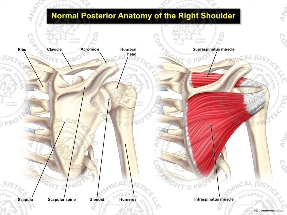
Normal Posterior Anatomy of the Right Shoulder from anatomicaljustice.com • both the circumflex arteries form an anastomosing circle around the surgical neck of. .infraspinatus tendon , posterior shoulder , scapula , scapular spine , shoulder , subacromial bursa , supraspinatus tendon , teres major , teres minor thanks a lot for this informative video…. Make anatomy really easy to learn…. • review pertinent anatomy and pathology associated with common causes of shoulder pain. The tendon of the infraspinatus passes posteriorly on to the. In the shoulder, articular cartilage covers the end of the humerus and socket area of the glenoid on the scapula. Specifically, the four rotator cuff muscles include the following Just below the anatomic neck are the greater and lesser tuberosities, where the muscles of the rotator cuff attach to.
Infrspinatus tendon and teres minor.
Posterior band of the ighl. Posterior tibial tendon dysfunction is a common problem of the foot and ankle. Posterior — the back of the shoulder. Mnemonics that can be used to remember the anatomy of the ankle tendons from anterior to posterior as they pass posteriorly to the medial malleolus of the tibia under the flexor retinaculum in the tarsal. Secondary restaint to inferior translation in the abducted shoulder. Shoulder anatomy for ultrasound evaluation. Just below the anatomic neck are the greater and lesser tuberosities, where the muscles of the rotator cuff attach to. Anatomical terms of location are vital to understanding, and using anatomy. • the anterior & posterior circumflex humeral artery. Using mr arthrography, we examined normal anatomy, anatomic variations, and pitfalls of imaging the labral capsular. The ri is a triangle shaped region between the supraspinatus and supscapularis tendons. Back (posterior) muscles of the shoulder. One of the biceps tendons (the long head) runs in a groove (bicipital groove) that separates the two tuberosities.
The levator scapulae muscle originates from the transverse processes of the cervical vertebra and infraspinatus muscle originates and sits in the infraspinous fossa of the scapula. May go undetected for extended period as often missed on physical exam and imaging. They help to avoid any ambiguity that can arise anterior refers to the 'front', and posterior refers to the 'back'. Secondary restaint to inferior translation in the abducted shoulder. Tendon pathology most commonly progresses posteriorly to the infraspinatus.
Shoulder Tendon Anatomy / The Shoulder - Anatomy,Function ... from image.slideserve.com Posterior tibial tendon (ptt) lies posterior to the medial malleolus before dividing into 3 limbs. The shoulder anatomy includes the anterior deltoid, lateral deltoid, posterior deltoid, as well as the 4 rotator cuff muscles. Presence of deep posterior shoulder pain. Tendon pathology most commonly progresses posteriorly to the infraspinatus. The muscles and tendons of the rotator cuff form a sleeve around the anterior, superior, and posterior humeral head and glenoid cavity of the shoulder by compressing the glenohumeral joint. Make anatomy really easy to learn…. Upper limb, breast, posterior shoulder, lateral chest wall. Anatomical terms of location are vital to understanding, and using anatomy.
• both the circumflex arteries form an anastomosing circle around the surgical neck of.
Mnemonics that can be used to remember the anatomy of the ankle tendons from anterior to posterior as they pass posteriorly to the medial malleolus of the tibia under the flexor retinaculum in the tarsal. The supraspinatus tendon is the most commonly affected tendon in the rotator cuff. Classically associated with seizures and lightning strikes. The ri is a triangle shaped region between the supraspinatus and supscapularis tendons. Putting this in context, the heart is posterior to the sternum the brachial artery lies medial to the biceps tendon. Inserts onto navicular tuberosity and first cuneiform. Back (posterior) muscles of the shoulder. Anterior graphic of the shoulder. Infraspinatus and teres minor tendon. Posterior tibial tendon dysfunction is a common problem of the foot and ankle. Learn vocabulary, terms and more with flashcards, games and other study tools. Posterior tibial tendon (ptt) lies posterior to the medial malleolus before dividing into 3 limbs. Related online courses on physioplus.
Specifically, the four rotator cuff muscles include the following The human shoulder is made up of three bones: Prevents anterior and posterior translations of the humeral head at greater degrees of abduction. An image depicting shoulder anatomy can be seen below. The supraspinatus tendon and subacromial bursa).
Posterior Shoulder Muscle Anatomy | Shoulder muscle ... from i.pinimg.com Tendon pathology most commonly progresses posteriorly to the infraspinatus. Infrspinatus tendon and teres minor. Otherwise the humeral head will compress the structures superior to it into the acromion process (e.g. Can lead to rupture of one or more of the tendons of the muscles forming the rotator cuff; .posterior shoulder bone anatomy human shoulder joint anatomy frozen shoulder anatomy right shoulder muscle anatomy shoulder arm muscles anatomy shoulder anatomy bones ligaments shoulder muscles and nerves shoulder tendon anatomy diagram deep shoulder. Assoc prof craig hacking ◉ ◈ and dr jeremy jones ◉ et al. The supraspinatus tendon is the most commonly affected tendon in the rotator cuff. Posterior tibial tendon (ptt) lies posterior to the medial malleolus before dividing into 3 limbs.
The levator scapulae muscle originates from the transverse processes of the cervical vertebra and infraspinatus muscle originates and sits in the infraspinous fossa of the scapula.
Being an undergraduate student excites me and inspires me to lean. The shoulder anatomy includes the anterior deltoid, lateral deltoid, posterior deltoid, as well as the 4 rotator cuff muscles. Tendon pathology most commonly progresses posteriorly to the infraspinatus. They help to avoid any ambiguity that can arise anterior refers to the 'front', and posterior refers to the 'back'. Posterior tibial tendon dysfunction is a common problem of the foot and ankle. • the anterior & posterior circumflex humeral artery. A tear in the rotator cuff may. Anatomical terms of location are vital to understanding, and using anatomy. The supraspinatus tendon and subacromial bursa). Anatomy of the suprascapular nerve. • review imaging findings relevant to these causes of pain and discuss a rationale for appropriate use. Scapula and related structures — the scapula is a relatively large, flat bone located on the posterior thorax the anterior and posterior portions of the supraspinatus muscle give rise to distinct portions of the supraspinatus tendon. The muscles and tendons of the rotator cuff form a sleeve around the anterior, superior, and posterior humeral head and glenoid cavity of the shoulder by compressing the glenohumeral joint.
Upper limb, breast, posterior shoulder, lateral chest wall shoulder tendon anatomy. In the shoulder, articular cartilage covers the end of the humerus and socket area of the glenoid on the scapula.


0 Comments:
Posting Komentar